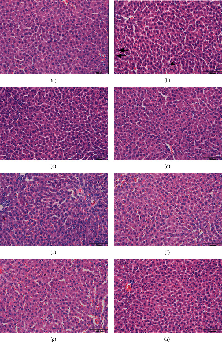
Figure 2.

Light microscopic images of liver tissue. (a) Control, (b) cis, (c) gal, (d) sly, (e) gal + sly, (f) cis + gal, (g) cis + sly, and (h) cis + gal + sly. Straight arrow: pyknotic hepatocytes. Dashed arrow: karyolytic hepatocytes. Arrowhead: Kupffer cell.
