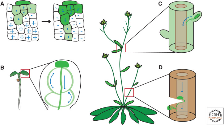
Figure 4.
Canalization processes for vasculature formation and regeneration. (A) Auxin source (dark green color) polarizes originally homogenous cells to create a directional transport (blue arrows) away from the source. The self-organizing property of auxin transport allows to canalize auxin from an initially broad domain into a narrow channel with high auxin-transporting capacity. (B) Auxin maximum in a cotyledon tip drives auxin canalization in a conserved pattern, demarcating the position of future vasculature. Transport-independent patterning mechanisms are likely also involved (Verna et al. 2019; for review, see Lavania et al. 2021). (C) The shoot apex is a well-known source of auxin, which keeps lateral buds inhibited. Once the apex is removed, the closest lateral bud is released from the inhibition and becomes a new dominant auxin source. Auxin canalization from this bud guides vasculature formation, connecting the lateral bud to the preexisting stem vasculature (Balla et al. 2011). (D) Wounding of stem vasculature results in a local auxin accumulation above the wound. Subsequently, auxin is canalized around the wound to initiate reconnection of the preexisting vasculature (Sauer et al. 2006; Mazur et al. 2016). Local external auxin application to the side of the stem also triggers auxin channel formation and guides formation of vasculature (Mazur et al. 2020b). Other cases of auxin canalization are during organogenesis at the shoot apical meristem and during embryogenesis (see Fig. 2).

