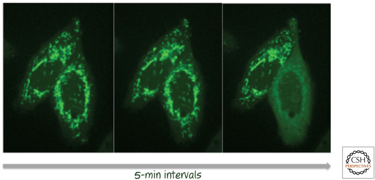
Figure 6.
Mitochondrial outer membrane permeabilization (MOMP). Cells expressing a fusion between cytochrome c and green fluorescent protein (GFP) undergoing MOMP in response to an apoptosis-inducing stress (left to right). As MOMP occurs (cell on the right in each pair), the distribution of fluorescence changes from localization in the mitochondria to being diffuse throughout the cytoplasm. (The cell to the left underwent MOMP at a later time.) The time between images is 5 min, with the first image taken several hours after the initial stress. (Images provided by Dr. Stephen Tait, St. Jude Children's Research Hospital, Memphis, Tennessee.)

