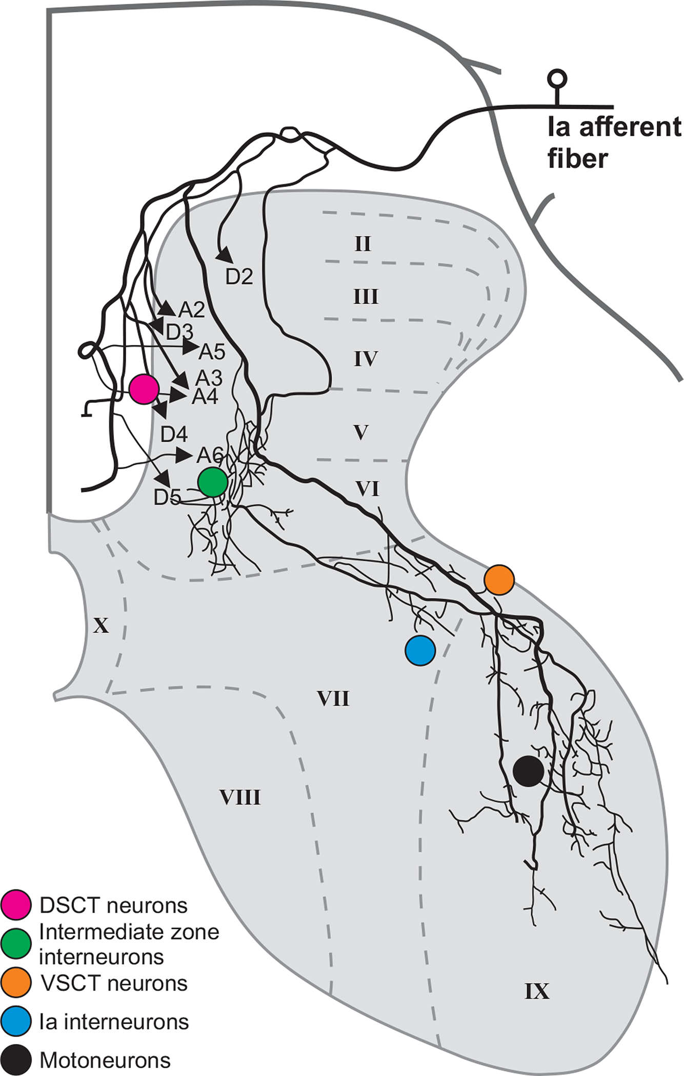
Figure 6. Projections from single group Ia afferent to neuronal targets of spinal cord.

The figure shows the trajectory of axon collaterals of a muscle spindle primary afferent (group Ia) from the medial gastrocnemius intra-axonally labelled with horseradish peroxidase. Colored circles indicate the location of five populations of target cells contacted by terminal branches of the group Ia afferent. Approximate locations of spinal cord laminae are shown (from Roman numerals II to X). Adapted and reproduced with permission from (435) using material from (419). DSCT, dorsal spinocerebellar tract; VSCT, ventral spinocerebellar tract).
