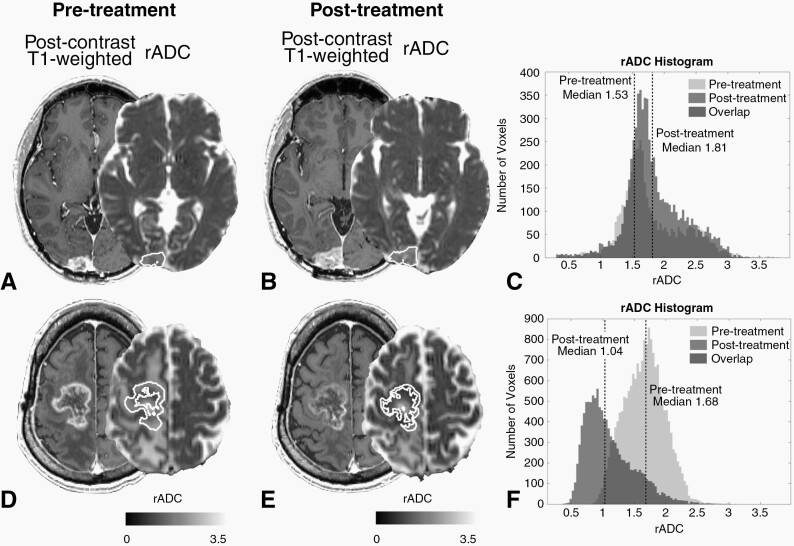
Fig. 2.
Postcontrast T1-weighted images and rADC maps of two representative patients with GBM who were treated with ICIs. Contrast-enhancing tumor volumes of interest are outlined in white. (A–C) The (A) pre- and (B) postscans of a 51-year-old male patient who showed an increase in both tumor volume and median rADC after ICI treatment (+73.2% and +8.9%, respectively; rADC histograms are shown in (C)). Although the tumor volume increased after treatment, the patient’s post-treatment rADC was relatively high (1.81) and OS was long (22.5 months). (D–F) The (D) pre- and (E) postscans of a 65-year-old male patient who showed a decrease in both tumor volume and median rADC after ICI treatment (-37.6% and -38.2%, respectively; rADC histograms are shown in (F)). Although the tumor volume decreased after treatment, the patient’s post-treatment rADC was relatively low (1.01) and OS was short (1.0 month).

