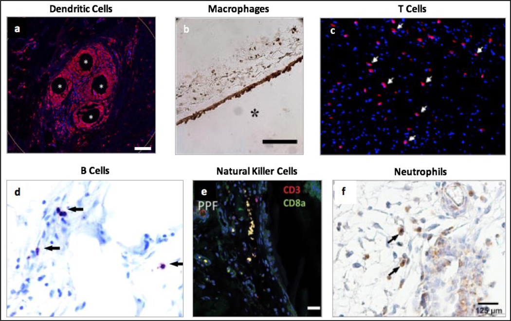
Figure 1.
Different cell types in the foreign body reaction to implanted polymeric biomaterials. a) Presence of dendritic cells shown by CD11b (red) staining of tissue samples from humans, approximately 1 year after surgery to repair an abdominal wall hernia using polypropylene mesh. Scale bar = 100 μm. Reprinted from Dievernich et al. Hernia, 2021 under the terms of the Creative Commons CC-BY License [13]. b) Presence of macrophages shown by mac3 (brown) staining of tissue samples from immunocompetent C57BL/6 mice, 4 weeks after a subcutaneous implantation of a poly-ethylene glycol hydrogel. Scale bar = 100 μm. Reprinted with permission from Lynn et al. J Biomed Mater Res A, 2011, 96: 621–631 (John Wiley & Sons) [14]. c) Presence of T cells shown by CD3 (red) staining of tissue samples from Sprague-Dawley rats, 3 weeks after a subcutaneous implantation of a polyurethane-encapsulated biosensor. Reprinted with permission from Ward et al. J Biomater Sci Polym Ed, 2008, 19: 1065–1072 (Taylor & Francis) [15]. d) Presence of B cells shown by B220 (purple) staining of tissue samples from specific pathogen-free female C57BL/6 mice, 4 weeks after a subcutaneous materials injection of nylon mesh. Reprinted with permission from Higgins et al. Am J Pathol, 2009, 175: 161–170 (Elsevier) [16]. e) Presence of natural killer cells shown by CD8a (green) staining of tissue samples from immunocompetent Sprague-Dawley rats, 6 weeks after an intradermal implantation of a polypropylene fumarate coated implant. Scale bar = 20 μm. Reprinted with permission from Bracaglia et al. J Biomed Mater Res A, 2019, 107: 494–504 (John Wiley & Sons) [17]. f) Presence of neutrophils shown by anti- myeloperoxidase antibody (brown) staining of tissue samples from C57BL/6 mice, 14 days after implantation of Dacron protheses in striated muscle tissue. Reprinted with permission from Moussavian et al. J Vasc Surgery, 2016, 64: 1815–1824 (Elsevier) [18].

