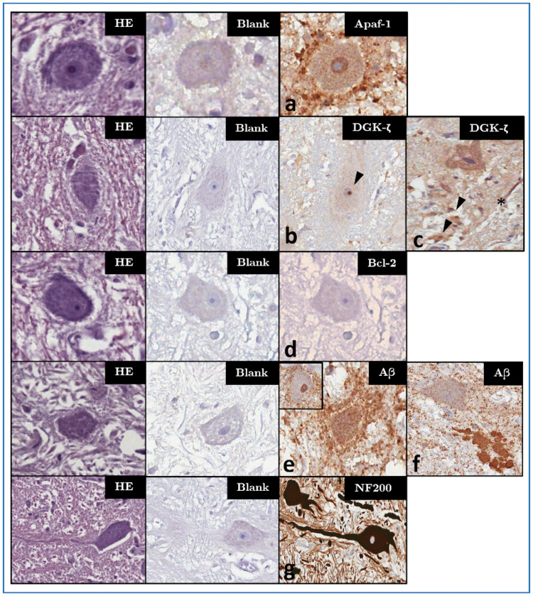
Fig 2. Haematoxylin-eosin/HE (left column), blank control/CTRL (middle column) and IHC patterns (right columns) for the investigated markers—magnification: 400x.
Panels marked a-f represent characteristic IHC patterns for each marker: (a) Apaf-1-IR in both neuronal cytoplasm and, unlike in most other examined animals, in the nucleus of ID319. (b) Nucleolar DGK-ζ-IR (arrowhead) in the VCN of a younger adult dolphin without brain lesions (ID146) as compared to (c) its cytoplasmic appearance in the VCN of a bycaught, older adult (ID203). In this animal, multifocal cytoplasmic IR in glial cells (arrowheads), as well as streaks of IR in the neuropil (asterisk) are apparent. (d) Lack of Bcl-2-IR in the VCN of a younger adult dolphin (ID89), representative of most adults. (e) Perineuronal enhancement of glial Aβ-IR in the VCN of a bycaught, hypoxic dolphin (ID203). (f) Aβ-IR marking a coalescing extracellular plaque between the VCN and superior olivary complex of older adult (ID319). An adjacent neuron shows light disseminated granular IR in its cytoplasm, but not in the nucleus, as in the VCN of most investigated adult dolphins <30 y.o. (inset in e). (g) Intense NF200-IR of the dendritosomatic and axonal cytoskeleton of VCN neurons (ID89, representative of all investigated animals).

