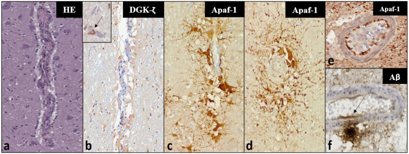
Fig 4. Histochemical (a) and IHC (b-f) findings in vascular and perivascular tissues in the investigated dolphins—Magnification: 100x.
Panels a-c represent the same vessel in consecutive sections. (a) Microscopic appearance of a mid-caliber vessel in the IC of ID344 using a haematoxylin-eosin staining, with a mild perivascular infiltrate characterized by mononuclear inflammatory cells. (b) very mild cytoplasmic DGK-ζ-IR in few foci of perivascular glia. Inset: Moderate, multifocal IR in pericapillary neuropil of ID133 (arrow)—magnification: 200x. (c) The same vessel of ID344 displays intense Apaf-1-IR, mostly in the cytoplasm of pericytes, showing perivascular distribution and radiating cellular processes, visible also in transverse sections of the IC vessels of ID344 (d). (e) Clearly demarcated endothelial Apaf-1-IR in a VCN vessel of ID142. (f) Aβ-IR deposit in neuropil adjacent to a mid-caliber VCN vessel—the IR appears to extend into the vessel wall and endothelial lining (arrow).

