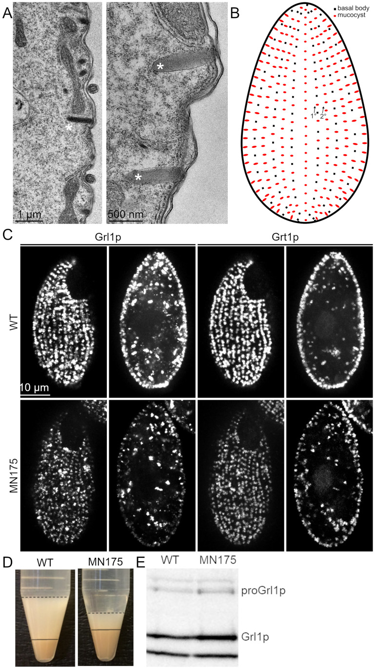
Fig 1. Mucocyst accumulation, docking and secretion.
A. Thin section electron micrographs of mucocysts in WT cells. The large majority of mucocysts (marked with *) are docked at the plasma membrane. The electron-dense contents are organized as a protein crystal, more easily seen at high magnification (right panel). Scale bars are indicated. B. Cartoon of a Tetrahymena cell, highlighting that the docked mucocysts (red) are aligned along cytoskeletal ‘ribs’ called 1°and 2°meridians. On the former but not the latter, mucocysts are interspersed with cilia, whose basal bodies (black) are shown. C. Mucocyst visualization in wildtype cells and MN175 cells. Wildtype cells and MN175 cells were starved for 3 hrs, fixed, permeabilized and immunolabeled with antibodies against mucocyst core proteins Grt1p (which localizes to one pole of the mucocyst core) and Grl1p (which localizes throughout the mucocyst core) (4D11 and anti-Grl1p, respectively). Surface and cross-sectional images were captured with Marianas Yokogawa type spinning disk inverted confocal microscope, 100X). Wildtype and MN175 cells were indistinguishable, both showing mucocysts with the expected distribution of Grl1p and Grt1p, docked at regular intervals along meridians. Scale bar is shown. Mucocyst docking was quantitatively analyzed by calculating the fraction of total Grl1p signal intensity in cross-sectional images that is present at the cell periphery, as described in Materials and Methods. Wildtype vs. MN175 cells showed no quantitative difference in mucocyst docking: wildtype = 23%; MN175 = 23% (n = 15, no statistically significant difference by Anova: Single Factor). D. MN175 cells are defective in induced mucocyst secretion. Mucocyst exocytosis was triggered in wildtype and MN175 cells by exposing them briefly to the calcium ionophore, dibucaine. Following centrifugation, the mucocyst contents released from the cells were quantified based on the volume of the flocculent layer (dashed line) above the packed cell pellet (solid line). The relatively small flocculent layer produced by MN175 cells indicates a defect in mucocyst secretion. The wildtype cell pellet is smaller, because a larger fraction of the wildtype cells remain trapped in the flocculent layer. E. MN175 accumulates higher levels of mucocyst protein. Resolved whole cell lysates of wildtype and MN175 cells (104 cells/4-20% SDS-PAGE lane) were immunoblotted with anti-Grl1p. Compared to wildtype cells, MN175 shows approximately 35% increased levels of mature Grl1p as well as approximately 25% increased levels of the unprocessed precursor, proGrl1p. The bottom band in each lane represents a cross-reactive species that shows no change from wildtype to MN175, and provides a loading control.

