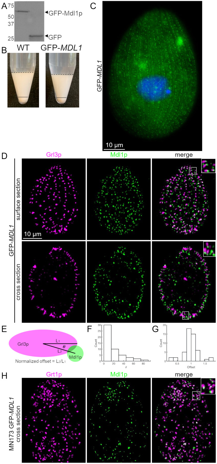
Fig 3. Mdl1p localizes to mucocyst tips.
A. GFP-Mdl1p was immunoprecipitated from cryomilled extracts of cells expressing the transgene at the MDL1 locus. Immunoprecipitates were resolved by SDS-PAGE and immunoblotted with anti-GFP. As shown in the left lane, GFP-Mdl1p appears as a single band of MW ~65 kDa (based on 2 gels). The positions of MW standards are shown on the left, and GFP by itself is shown in the right lane. B. Dibucaine stimulation of mucocyst exocytosis. Cells in which all copies of MDL1 are GFP-tagged show wildtype levels of mucocyst secretion upon stimulation with dibucaine, indicating that tagged Mdl1p retains activity. C. Live cell imaging of GFP-Mdl1p. Cells expressing GFP-Mdl1p at the endogenous locus were grown to stationary phase, fixed, and the nuclei stained with DAPI (blue). As shown here for an individual cell, small green fluorescent puncta extend in a linear array aligned with the long axis of the cell with spacing expected for docked mucocysts at 1° and 2° meridians, as explained in the text. The larger and less distinct green signals within the cytoplasm are due to autofluorescence from mitochondria and food vacuoles. Live immobilized cells were imaged using a Zeiss Axio Observer 7 microscope, with objective lens 100X. Scale bar is indicated. D. Polar distribution of Mdl1p on mucocysts. Cells expressing GFP-Mdl1p were fixed and immunolabeled with DyLight 650-conjugated antibodies against the mucocyst core protein Grl3p. In these panels, the GFP signal is colored green, and the DyLight signal is colored fuchsia. GFP-Mdl1p is concentrated at the tips of mucocyst tips where they dock at the plasma membrane, most clearly seen in the merged image inserts (right-most images). Tangential and cross sections of individual cells are shown. Scale bar is indicated. E. An explanatory cartoon depicting the angles in F and distance (offset) in G. F. Test for tip localization: histogram of the angles (°) made by intersecting the line passing through the centroids of overlapping Grl3p and GFP-Mdl1p speckles with the long axis of the Grl3p speckle. 0° indicates perfect alignment of the GFP-Mdl1p speckle along the long axis of the Grl3p speckle, while 90° indicates perfect alignment along the short axis. G. Histogram of the distances between the centroids of overlapping Grl3p and GPF-Mdl1p speckles, normalized to half the length of the Grl3p speckle (n = 32 speckles from 6 cells). H. Distribution of Mdl1p on non-docked mucocysts. MN173 mutant cells, in which mucocysts fail to dock, were transformed to express GFP-Mdl1p at the MDL1 locus. Overnight cultures were fixed and immunolabeled with antibodies against the mucocyst tip protein Grt1p, which is distributed broadly at the docking end of the mucocyst. Because the mucocysts are large and present in random orientations, only a small number of tips appear in each optical section. Many mucocysts show clear polarization, with GFP-Mdl1p at one tip. Cells were imaged using Marianas Yokogawa type spinning disk inverted confocal microscope, 100X. Ilastik (pixel classification software) was used to filter out diffuse autofluorescence arising from mitochondria.

