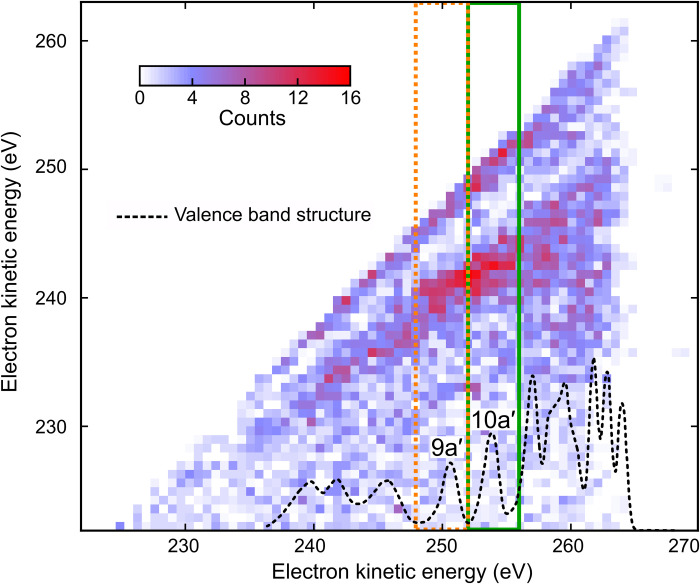
Fig. 5. Two-electron coincidence spectroscopy of glycine with 274.0-eV photons.
Coincidence map of two-electron detection. Photoelectron (10a′ and 9a′)–Auger electron coincidences following the resonant x-ray transition (1s → 1h) are indicated (green and orange squares). The density of valence states is indicated (dotted black) by using the experimental data reproduced from (14). Valence electron emission from x-ray probe-induced sequential double ionization contributes to an uncorrelated background with respect to the (initial) x-ray pump-induced photoionization events, which allows observing the fingerprint of the probe-induced Auger decay. The map also shows an enhanced probability for the detection of two (photo)electrons with an equal kinetic energy, which are the likely result of a false coincidence between two valence ionization events.

