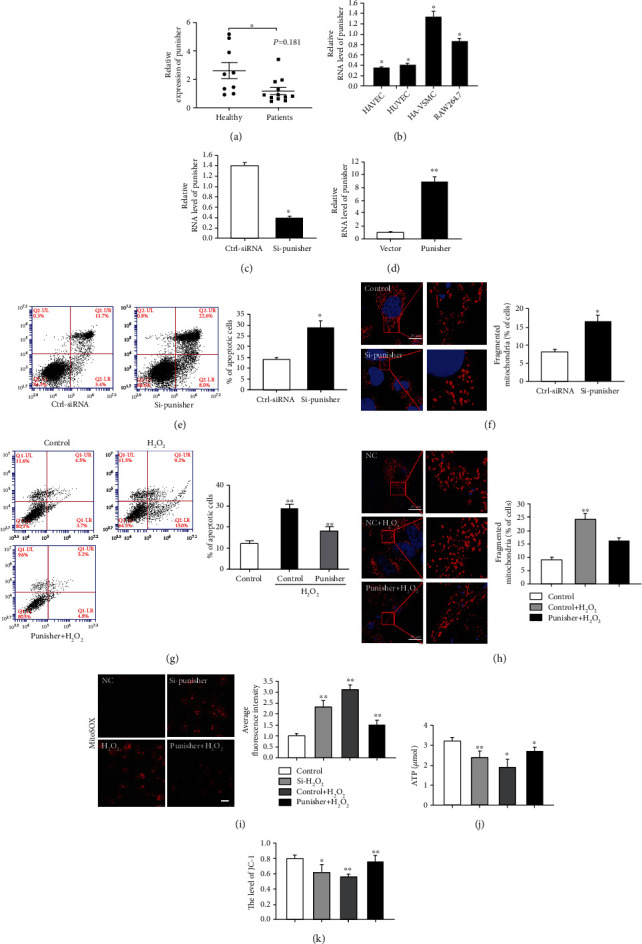
Figure 1.

Punisher regulates cell apoptosis and mitochondria dynamics of VSMC. (a) Punisher transcript expression in atherosclerotic plaques of patients (n = 12) and healthy people (n = 9) was measured by quantitative reverse transcription–polymerase chain reaction (qRT-PCR). (b) qRT-PCR analysis of Punisher mRNA expression in human arterial vascular smooth muscle cells (HA-VSMC), RAW264.7, and endothelial cells. (c, d) siRNAs and overexpression plasmids were designed and transfected for 24 hours to knock down or enforce Punisher expression in VSMCs. The expression of Punisher was quantified by qRT-PCR. (e) Annexin V-FITC/propidium iodide staining and FACS quantification of the number of apoptotic cells in HA-VSMC after transfection of si-Punisher. (f) MitoTracker staining of mitochondrial fission in VSMC after Punisher knockdown. (g) Annexin V-FITC/propidium iodide staining and FACS quantification of the number of apoptotic cells in HA-VSMC after transfection of Punisher overexpression plasmids with H2O2 stimulation. (h) MitoTracker staining of mitochondrial fission in VSMCs after Punisher overexpression with H2O2 stimulation. (i, j) Evaluation of ROS and ATP production after knockdown or overexpression of Punisher with or without H2O2 treatment. (k) Measurement of mitochondrial membrane potential by detecting mitochondrial membrane potential expression using JC-1. All values are the average of at least 3 biological replicates, and data shown are the mean ± SD. Scale bars: 20 μm. ∗p < 0.05; ∗∗p < 0.01.
