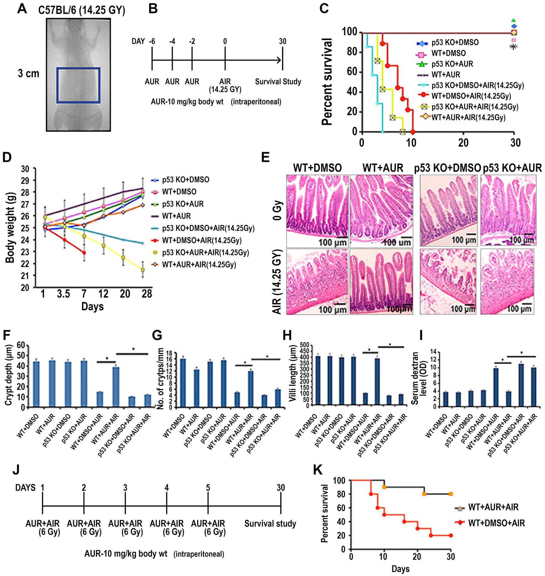
Figure 1:

Auranofin treatment protected C57BL/6 mice from radiation-induced lethality. A) Schematic diagram demonstrating AIR exposure field for C57BL/6 mice. A 3 cm square abdominal area of the mouse containing the GI tract was irradiated (irradiation field), thus shielding the upper thorax, head and neck as well as lower and upper extremities, protecting a significant portion of the bone marrow. Mice were treated with auranofin (10 mg/kg i.p.) on days 2, 4 and 6 prior to AIR (14.25 Gy). B) Schematic diagram of the treatment protocol. C) Kaplan–Meier survival analysis. Wild type (WT) male mice receiving auranofin showed 100% survival beyond 30 days post irradiation compared with mice receiving DMSO where 100% of mice died within 10 days after irradiation (p<0.0001, Log-rank (Mantel–Cox) test; n=10 per group). However, auranofin did not inhibit radiation-induced lethality in p53 knockout (KO) mice. D) Body weight of mice at times post irradiation (14.25 Gy AIR). E) Representative H&E stained sections of mouse jejunum (20X). Note, restitution of crypt villus structures in irradiated, auranofin treated WT mice compared with mice receiving only radiation. However, in p53 KO mice auranofin treatment did not protect the intestinal epithelium from radiation damage. F-H) Histograms showing the crypt depth (F), number of crypts/mm (G) and villous length (H) in jejunal sections of WT mice and mice treated with auranofin or AIR or AUR+AIR. Auranofin treatment did not have any effect on the intestine but significant increase in crypt depth (*, p<0.001), crypt count/mm (*, p<0.005) and villi length (*, p<0.001) were observed in the AIR cohort pre-treated with auranofin compared to the AIR only cohort (unpaired t-test, two-tailed) (n=6 per group). AUR treatment in p53 KO mice demonstrated significantly less crypt depth (*, p<0.002), crypt count/mm (*, p<0.006) and villous length (*, p<0.003) compared to WT mice receiving auranofin. I) Histogram demonstrating serum dextran levels in WT and p53KO mice. No significant change in serum dextran level was observed in control and in WT mice treated only with auranofin. Significantly higher serum dextran levels were observed in the AIR group compared to the AIR+AUR group (*, p<0.006) or control (DMSO) group (p<0.004) of WT mice. In p53KO mice AUR pre-treatment did not reduce the serum dextran level following irradiation. J) Schematic diagram of the treatment protocol and fractionated radiation schedule. K) Kaplan–Meier survival analysis of mice receiving fractionated AIR (6 Gy × 5 fraction). Auranofin pre-treatment improved survival in these mice compared to controls (p<0.0002, Log-rank (Mantel–Cox) test; n=10 per group).
