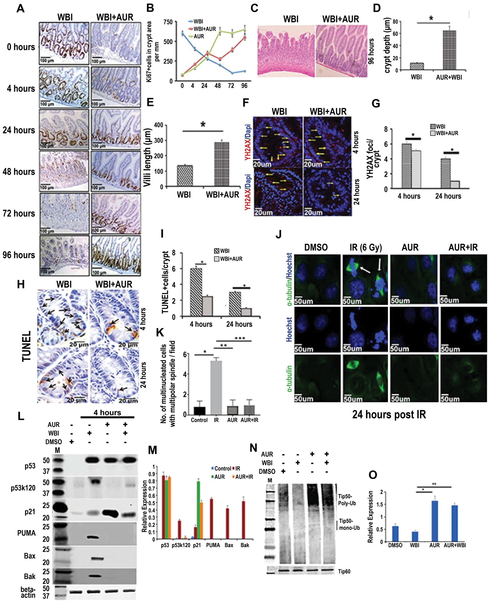
Figure 2:

Auranofin modulates crypt cell proliferation kinetics, reduces radiation-induced DNA damage and promotes crypt regeneration. C57BL/6 male mice pretreated with auranofin (10 mg/kg i.p.) or vehicle (DMSO) were exposed to 12.5 Gy WBI. A) Representative images of jejunal sections stained with Ki67. B) Quantification of Ki67 positive cells in jejunum from irradiated mice treated with auranofin or DMSO, as a function of time after irradiation. C) H&E stained jejunal section at 96 h post WBI (n=6 mice per group). D–E) Histogram demonstrating crypt depth and villous length in jejunal sections at 96 h post WBI. Auranofin pretreatment demonstrated higher crypt depth (p<0.0003) and villous length (p<0.0006) compared to WBI control. F) Representative confocal images of γH2AX staining of jejunal sections. G) Histogram demonstrating number of γH2AX positive foci in crypt cells. Auranofin pretreatment reduced number of γH2AX positive foci/crypt compared to WBI control, both at 4 and 24 h post exposure (*p<0.05 and *p<0.003, respectively). H) Representative image of TUNEL staining in jejunal sections. I) Histogram demonstrating number of TUNEL positive cells per crypt. Auranofin pretreatment reduced number of TUNEL positive cells/crypt compared to WBI control, both at 4 and 24 h post exposure (*p<0.008 and p<0.006, respectively). J. Representative confocal microscopic images of α-tubulin and Hoechst staining demonstrate mitotic catastrophe (α-tubulin and Hoechst double positive cells indicated with an arrow). K) Histogram of number of multinucleated cells with multipolar spindle per field (n=3 mice/group). Auranofin pretreatment reduced the number of these cells compared to the IR control (***p<0.0003). Immunoblot demonstrating expression of total p53, acetylated p53 (p53k120), total p21, PUMA, BAX and BAK in crypt cells. M) Histogram demonstrating densitometric analysis of immunoreactive bands representing expression of the above proteins (assessments repeated in 3 mice/group). Significant upregulation of p53k120 (p<7.86E-07), PUMA (p<7.96E-08), BAX (p<6.87E-08), BAK (p<6.92E-08) expression was noted in the WBI group compared to the WBI+AUR group. The WBI group also showed significantly higher expression of these molecules compared to untreated controls [p53k120 (p<8.78E-08), PUMA (p<8.66E-08), BAX (p<8.87E-08), BAK (p<8.32E-08)] or auranofin alone [p53k120 (p<6.78E-06), PUMA (p<7.66E-07), BAX (p<5.87E-07), BAK (p<8.32E-08)]. Expression of p21 was significantly higher for AUR+WBI compared to WBI (p<5.68E-05). N). Immunoblot demonstrating ubiquitinated and total Tip60 in crypt cells. O) Histogram of results from densitometric analysis of immunoreactive bands representing ubiquitinated Tip60. The ubiquitinated form of Tip60 was significantly higher in AUR+WBI compared to WBI mice (p<0.0004).
