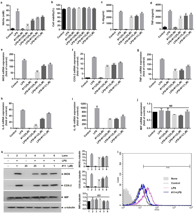
Fig. 8. Effects of compound 11 on the generation of pro-inflammatory factors in LPS-primed BV-2 microglial cells.
Cells were pretreated with compound 11 (1–50 μM) for 0.5 h prior to LPS (0.2 µg/mL) stimulation. a, b After 24 h of LPS stimulation, NO levels were evaluated by Griess reagent (a), and cell viability was determined by MTT assays (b). c, d The production of IL-6 (c) and TNF-α (d) in supernatants after LPS stimulation for 24 h was evaluated by ELISA. e–j The mRNA expression of iNOS, COX-2, TNF-α, IL-6, IL-1β, and MIF after LPS treatment for 6 h was assessed by RT-qPCR. k Western blot analysis of iNOS, COX-2, MIF, and α-tubulin protein expression in lysates of LPS-primed BV-2 microglial cells at 16 h (k, left). The expression of α-tubulin was used as an internal control, and the relative expression of MIF, iNOS, and COX-2 was quantified by densitometric analysis (k, right). l After 16 h of LPS stimulation, the production of intracellular ROS was measured by flow cytometry. *P < 0.05, **P < 0.01, ***P < 0.001 compared with the LPS-alone group.

