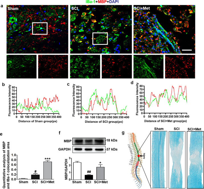
Fig. 5. Metformin enhances microglial cells to phagocytose myelin debris.
a Co-staining of IBa-1 (a specific protein of microglial cells) (red) and MBP (labeled for myelin) (green) in posterior horn area of spinal cord from each group at 14 d after SCI (n = 5). Scale bar = 25 μm. b–d Colocalization analysis of IBa-1 and MBP in selected area of spinal cord from each group. e Quantification analysis of MBP and IBa-1 colocalization area in co-staining of IBa-1 and MBP in each group at 14 d after SCI. f Western blotting and quantification analysis of MBP in the spinal cord from each group at 14 d after SCI (n = 3). g Luxol fast blue (LFB)-stained longitudinal sections of spinal cord from each group at 14 d post SCI (n = 5 per group). #P < 0.05, ##P < 0.01 vs. Sham group, *P < 0.05, ***P < 0.001 vs. SCI group. SCI spinal cord injury, Met metformin, MBP myelin basic protein.

