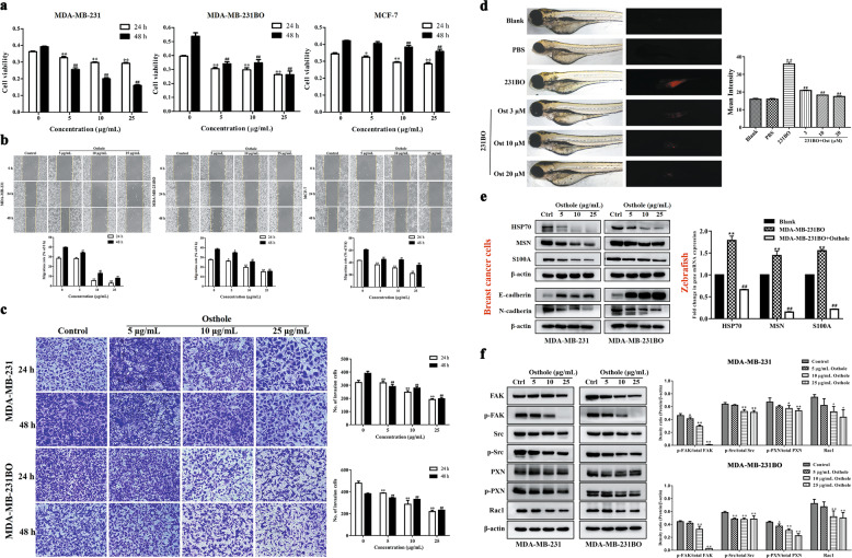
Fig. 2. Inhibitory effect of osthole at different concentrations on the proliferation, migration and invasion of highly metastatic breast cancer cells.
a–c After treatment with osthole (5, 10, and 25 μg/mL) for 24 and 48 h, the cell proliferation, migration and invasion in both MDA-MB-231 and MDA-MB-231BO cells were analyzed by SRB, wound-healing, and transwell assays, respectively. Data were expressed as the mean ± SD for triplicate. *P < 0.05, **P < 0.01 vs. the control group for 24 h; #P < 0.05, ##P < 0.01 vs. the control group for 48 h. Amplification factors: 100× for b and 200× for c. d A zebrafish breast cancer xenograft model was established by injecting Dil-labeled MDA-MB-231BO cells into the perivitelline cavity of each embryo. After treatment with different concentrations of osthole (3, 10, and 20 μmol/L) for 48 h, images of the disseminated tumor cells in the zebrafish body were captured using an inverted fluorescence microscope and the mean intensity of tumor cells was analyzed by ImageJ software. e Western blot was used to evaluate the effect of osthole on the expression of epithelial marker E-cadherin and the mesenchymal markers HSP70, MSN, S100A, and N-cadherin in highly metastatic breast cancer cells; real-time PCR was used to analyze the effect of osthole on the gene expression of HSP70, MSN, and S100A in the zebrafish breast cancer xenograft model. f Western blot was used to investigate the effect of osthole on the protein expression of FAK/Src/Rac1 signaling. Data were expressed as the mean ± SD for triplicate. **P < 0.01 vs. the blank group; ##P < 0.01 vs. the MDA-MB-231BO group. Ctrl: control; Ost: osthole.

