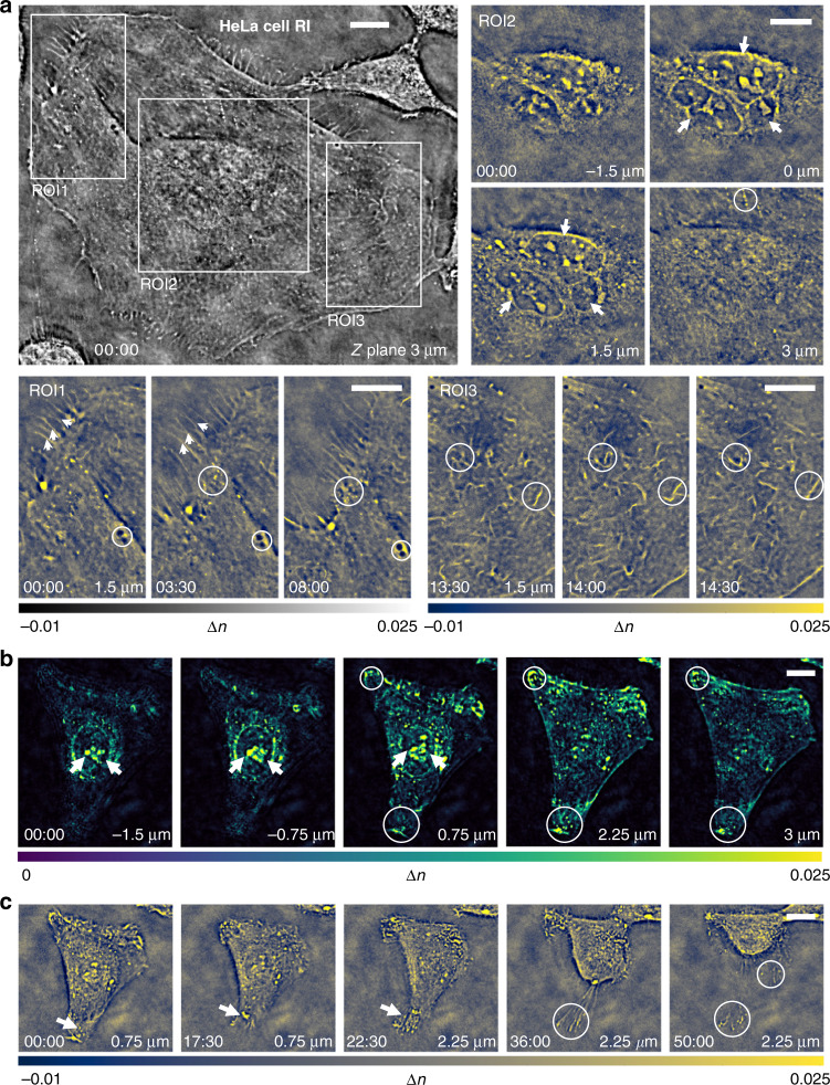
Fig. 5. HeLa cells 3D RI imaging over hour-long time-lapse.
a Recovered RI slice of trinucleated HeLa cell located at 3 µm Z plane at the start time point of 00:00:00, and enlarged time-lapse tomographic RI image of three different ROIs in the FOV. The whole process of HeLa cells visualization is given in Video S9. b Maximum intensity rendering of HeLa cell in another ROI located at different axial planes at the start time point of 00:00:00. c Cross-sectional view of the RI tomogram in the ROI at five different time points and axial planes. Scale bars: 15 µm.

