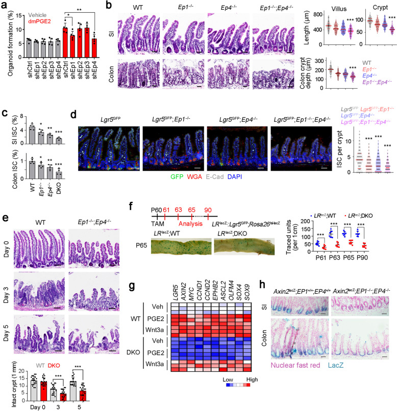
Fig. 7. PGE2 drives Wnt signaling activation to maintain ISC stemness.
a Organoid formation assay of indicated PGE2 receptor-silenced ISCs, supplemented with 1 μM dmPGE2. Results are from 5 independent experiments. b H&E staining of SI and colon of WT, Ep1−/−, Ep4−/−, and DKO mice. Typical images were shown in left panel and 100 fields from five mice were observed in right panel. Scale bars, 50 μm. c FACS detection for ISC ratios in SI and colon crypt, which were from WT, Ep1−/−, Ep4−/−, and DKO mice. 5 mice per group. d Fluorescence staining of intestinal tissue from 2-month-old Lgr5GFP, Lgr5GFP; Ep1−/−, Lgr5GFP; Ep4−/− and Lgr5GFP; Ep1−/−; Ep4−/− mice. 100 fields from five mice were observed. Scale bars, 50 μm. e WT and DKO mice were treated with 10 Gy’s radiation, and sacrificed at indicated time points, and typical images were shown. 20 fields from five mice for each group. Scale bars, 100 μm. f Lgr5GFP-CreERT2; Rosa26lsl-lacZ (LRlacZ) mice were crossed with DKO mice, and intestinal whole-mount staining of β-gal was performed for lineage tracing analysis. 8 mice were observed for each group and typical sections were shown. P60, postnatal day 60; TAM, tamoxifen. g Heatmap of indicated Wnt/β-catenin target genes in WT and DKO ISCs. 3 mice were used per group. h WT and DKO mice were crossed with Axin2lacZ mice. SI and colon tissues were stained for β-gal expression. β-gal staining indicates the activation of Wnt/β-catenin signaling. Typical images of 5 mice were shown. Scale bars, 50 μm. *P < 0.05, **P < 0.01, ***P < 0.001 by unpaired one-tailed Student’s t-test. At least three independent repeats were performed for each experiment and the representative results were shown.

