Figure 7.
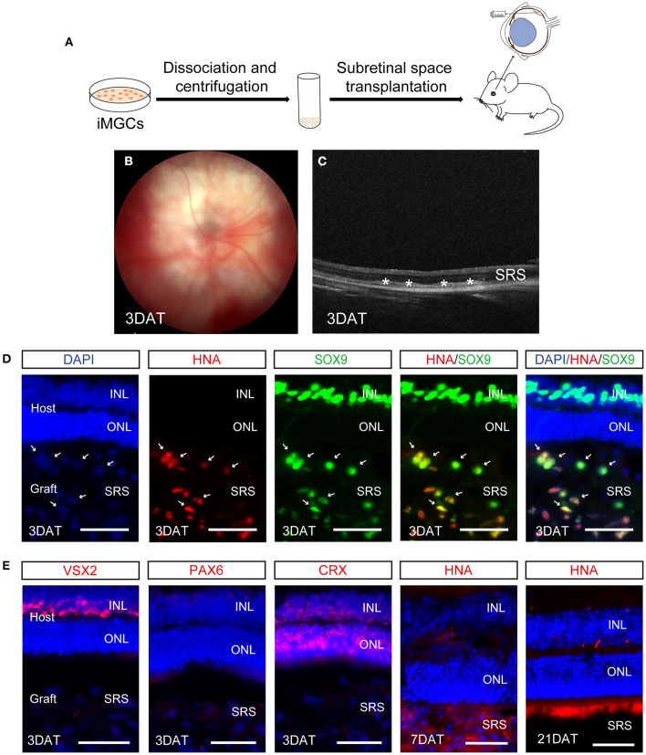
Evaluation of cell survival of the passaged iMGCs after subretinal transplantation into NOD/SCID mice. (A) Schematic overview of the subretinal transplantation process of the iMGCs. (B,C) Fundus photography (B) and optical coherence tomography (OCT) (C) showed the successful delivery of the iMGCs into subreinal space (SRS) in NOD/SCID mice 3 days after transplantation (DAT). *The grafted cells. (D,E) Immunofluorescence staining showed the grafted iMGCs (D3, P2 iMGCs from D120 BC1-ROs) were positive for HNA and SOX9, but negative for VSX2, PAX6, and CRX at 3 DAT, and gradually disappeared from 7 to 21 DAT. INL, inner nuclear layer; ONL, outer nuclear layer. Scale bars = 50 μm (D,E).

