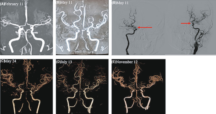
FIGURE 1.

Cerebral vascular imaging before and after this onset. (A) MRA before this onset was normal. (B) MRA on the 10th day of the onset without effective antibiotic therapy indicated the bilateral varying vascular stenosis and occlusion at the beginning of internal carotid artery and middle cerebral artery, posterior cerebral artery, and adjacent collateral blood vessel formation; digital subtraction angiography (DSA) indicated multiple localized stenosis in the intracranial segment of bilateral internal carotid artery, bilateral middle cerebral artery M1 segment, and the initial segment of the basilar artery. (C) CTA on the 24th day of the onset indicated the same vascular stenosis to b. (D) CTA after the 2 months of effective anti‐infective treatment showed the improvement of vascular stenosis. (E) CTA after half a year of effective anti‐infective treatment showed obvious vascular improvement and collateral circulation formation
