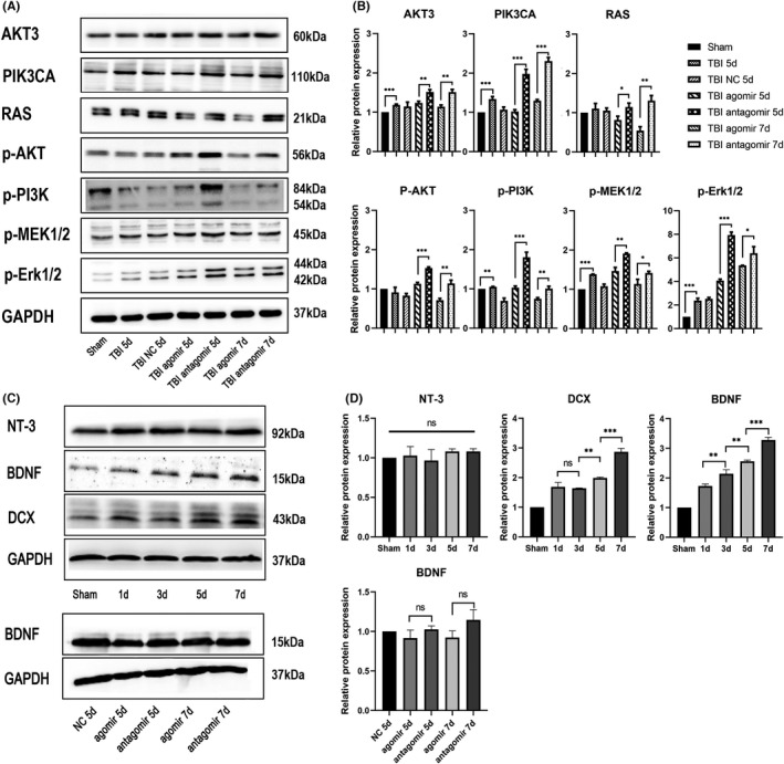
FIGURE 5.

Western blotting of SVZ tissues post‐TBI. (A) MiR‐124‐3p affected the expression of neurotrophin signaling pathway molecules after TBI. Western blot (WB) analysis of Akt3, PIK3CA, Ras, phospho‐Akt, phospho‐PI3K, phospho‐MEK1/2, and phospho‐Erk1/2 expression in SVZ tissues of TBI rats injected with agomir‐124‐3p or antagomir‐124‐3p 5 and 7 days after injury. (B) Statistical analysis of the relative protein expression of each molecule. All proteins analyzed were found to be significantly differentially expressed between the agomir group and antagomir group. (C) The expression of NT‐3, BDNF, and DCX in the SVZ tissue was tested 1, 3, 5, and 7 days after TBI. (D) Statistical analysis of the relative protein expression of each molecule. All data are presented as the means ± SD. ⁎ p < 0.05. n = 3/group. Full length western blot scans for the cropped images are presented in Figure S1
