FIGURE 2.
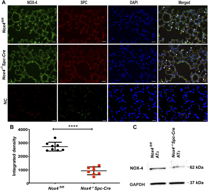
NOX4 expression is downregulated in the lungs and isolated AT2 cells from Nox4 −/− Spc-Cre mice. Nox +/- Spc-Cre animals were injected with tamoxifen to generate Nox4 −/− Spc-Cre animals and IHC was performed on the harvested lungs. (A) Representative immunofluorescent micrographs of lung tissue showing individual staining as well as co-staining of NOX4 and AT2 pneumocyte marker SPC (merge of green and red). Co-localization of NOX4 (green) and SPC (red) could be seen in lungs of Nox4 fl/fl mice, but not in Nox4 −/− Spc-Cre mice as indicated with white arrows (B) Quantified data of NOX4 fluorescence intensity from NOX4-SPC co-stained lung tissue (C) Expression of NOX4 in primary AT2 cells isolated from Nox4 fl/fl and Nox4 −/− Spc-Cre mice lung. n = 3 ****p < 0.0001.

