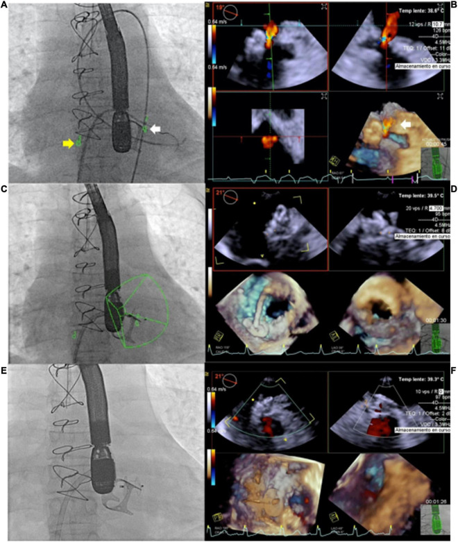
FIGURE 5.
Use of echocardiography-fluroroscopy fusion (EFF) to facilitate mitral paravalvular leak (PVL) closure. (A,B) Anatomical markers placed on fluoroscopy and echocardiography. Marker 1 (yellow arrow) indicate position of transeptal puncture. Marker 2 (white arrow) indicate site of mitral PVL. (B) Color flow Doppler confirming mitral PVL. Combined EFF image allows efficient crossing of PVL (C,D). Continuous feedback from EFF facilitates alignment of delivery sheath and device deployment. (E) Fluoroscopy showed good device position post release, (F) confirmed by echocardiography and no residual leak was observed. Courtesy of Dr. Juan Pablo Sandoval, Instituto Nacional de Cardiologia Ignacio Chavez, Mexico City, Mexico.

