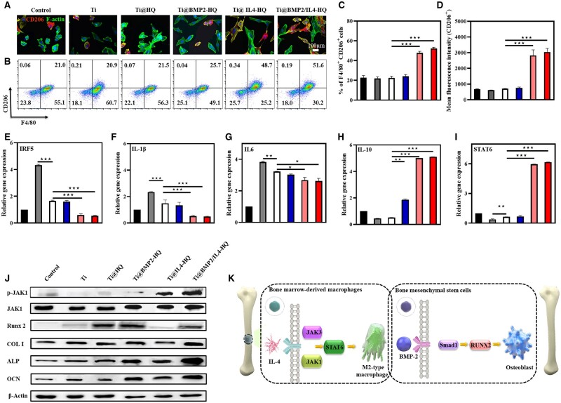
Figure 4.
Ti@HQ scaffold loaded with IL-4 effectively primed macrophages toward M2 phenotype. (A) Representative CLSM images of BMDMs treated for 48 h. Red channel, CD206; green channel, F-actin; blue channel, nucleus (n = 3). (B) Flow cytometry analysis of CD206 expression and the percentage of M2 type (C, F4/80+CD206+) macrophages. The gating approach for flow cytometry was based on FSC-A and SSC-A (Fig. S13) (n = 3). (D) Mean fluorescence intensity of CD206 staining (n = 3). (E–I) Real-time PCR of M1 polarization-related IRF5 (E), IL-1β (F) and IL-6 (G) and M2 polarization-related IL-10 (H) and STAT6 (I) (n = 3). (J) Protein expression level of p-JAK1, and JAK1 in macrophages runx-2, COL I, ALP and OCN in hBMSCs determined by western blotting. (K) The mechanism of M2-type macrophage polarization and osteogenic gene activation by Ti@BMP2/IL4-HQ. Data are shown as mean ± SDs (n = 5). **P < 0.01, ***P < 0.001, between the indicated groups

