
Fig. 4 |. Two-cell-signature genes are involved in telomere protection of ES cells.
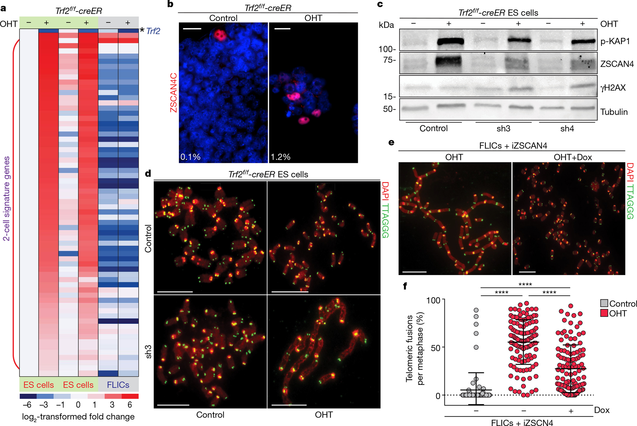
a, Heat map displaying the expression levels of 2-cell-signature genes between control and OHT-treated ES cells (two independent cell lines) and FLICs (one cell line). Expression levels are normalized to the values of the control ES cell line. b, Representative immunofluorescence staining for ZSCAN4C in control and OHT-treated ES cells. Percentages of ZSCAN4-positive cells are shown in bottom left corner. c, Western blotting analysis of control and OHT-treated ES cells expressing either a control shRNA or two independent shRNAs against Zscan4c (sh3 and sh4). d, Metaphase spreads derived from ES cells of the indicated genotypes and treatment. Additional information is provided in Supplementary Table 1, Source Data for Fig. 4. e, Representative metaphases derived from doxycycline-inducible-ZSCAN4 (iZSCAN4) FLICs, treated with doxycycline (+Dox) or not, were collected 4 days after treatment with OHT. f, Scatter plot representing the distribution of telomeric fusions. Each dot represents the percentage of telomeres fused in one metaphase spread; for detailed information, see Supplementary Table 1 and Source Data for Fig. 4. All data panels in the figure are representative of at least three experiments. NS, non-significant (P ≥ 0.05), P < 0.05, ****P < 0.0001, one-way ANOVA.