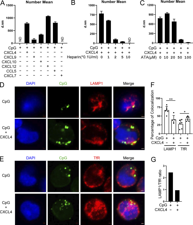
Figure 7.
Chemokines bind DNA to form nanoparticles, which are retained in the early endosomes of pDCs. (A) The nanoparticle size (number mean) of CpG DNA (0.1 μM) and the indicated combinations of chemokines (10 μg/ml) + CpG, in PBS were measured by DLS. All represented samples met the quality control criteria; otherwise the samples were annotated as ND. (B and C) The nanoparticle size (number mean) measurement of CXCL4 (10 μg/ml) and CpG DNA (0.1 μM) with or without the indicated concentration of heparin (B) or ATA (C) by DLS. (D–G) pDCs were cultured with 10 μM CpG-AF488, alone or with 10 μg/ml CXCL4 and 0.25 μM CpG-AF488, for 3 h. Representative confocal images of costaining of late endosome marker LAMP1 and CpG-AF488 (D) and early endosome marker TfR and CpG-AF488 (E) are shown. (F) Percentage of colocalization between CpG-AF488 and LAMP1+ or TfR+ endosome in CpG-AF488+ cells; 50–200 cells were evaluated in a blinded fashion for each donor (n = 5–6). (G) Ratio of CpG-AF488 localization in late endosome versus early endosome (LAMP1/TfR) in CpG-AF488 and CpG-AF488 + CXCL4–stimulated pDCs. All results are represented as means ± SEM. Statistical significance was evaluated using Mann–Whitney U test, and only comparisons that are significant are shown. *, P < 0.05; **, P < 0.01. All scale bars are 1 μm. d.nm, diameter in nanometers.

