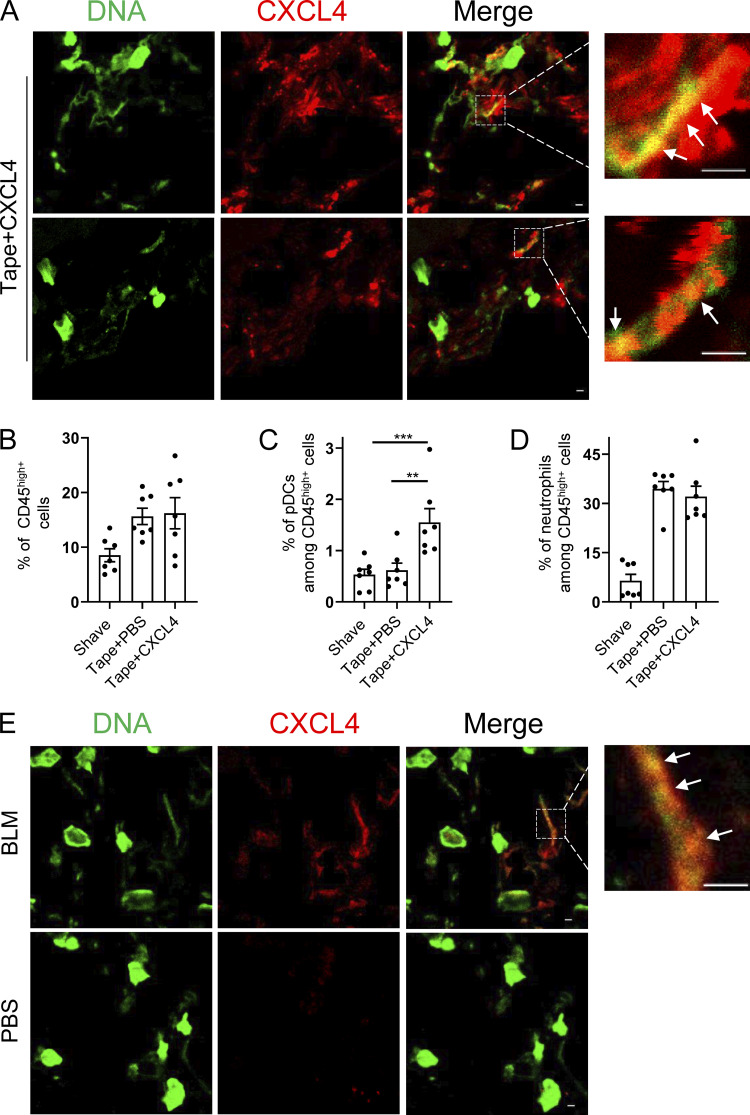
Figure 8.
CXCL4 can be associated with DNA in vivo during inflammatory responses in the skin. (A–D) Age-matched C57BL/6 mice were either shaved only or tape stripped (tape), followed 1 h later by an intradermal injection of CXCL4 (2 µg/mouse) or PBS. 6 h later, skin biopsies were collected, and CXCL4 and extracellular DNA (eDNA) were stained. (A) Representative confocal microscopy images of CXCL4 and eDNA colocalization. (B–D) Cellular skin infiltrates were characterized by flow cytometry and representative dot plots of CD45hi immune cells (B), CD45hiCD11b-Ly6G-Siglec H+ pDCs (C), and CD45hiCD11b+Ly6G+ neutrophils (D) are shown. All results are represented as means ± SEM from two independent experiments (n = 6–7 per group), and statistical significance was evaluated using Mann–Whitney U test; only comparisons that are significant are shown. **, P < 0.01; ***, P < 0.001. (E) Skin fibrosis was induced in age-matched C57BL/6 mice by s.c. daily injection of PBS (control) or BLM for 3 wk. Skin biopsies were collected, and CXCL4 and eDNA were stained. Representative confocal images of CXCL4 and eDNA colocalization are shown, and white arrows in A and E indicate colocalization. All scale bars are 2 μm.

