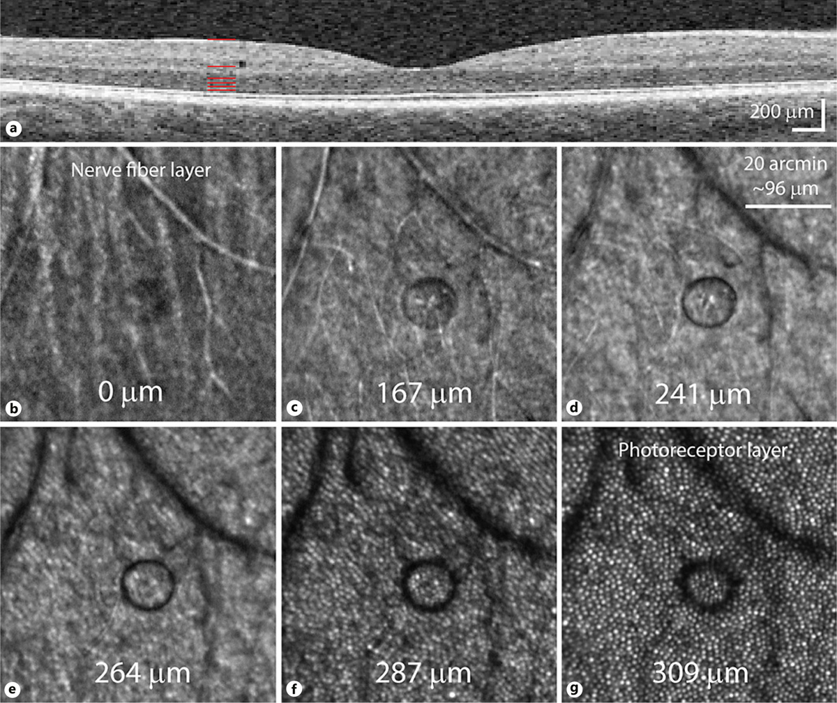
Fig. 2.

Microcyst appearance in different focal planes. a OCT scan taken 1 day after microcyst discovery, showing its location in the inner nuclear layer. Pixels are scaled to be isotropic. Red lines depict the corresponding depths of the AOSLO images in b–g, with depths estimated from the defocus used during capture (photoreceptors appeared sharpest at one depth, assumed to be near the ELM, that was calculated to be 309 μm below the nerve fiber layer image). b The nerve fiber layer shows a hyporeflective microcyst area. The microcyst border appears distinctly at 167 μm depth (c), becoming darker at 241 μm (d). As the imaging plane moves toward the photoreceptor layers (e–g), the microcyst becomes less well-defined and darker still, as it deflects more light away from the AOSLO light collection path.
