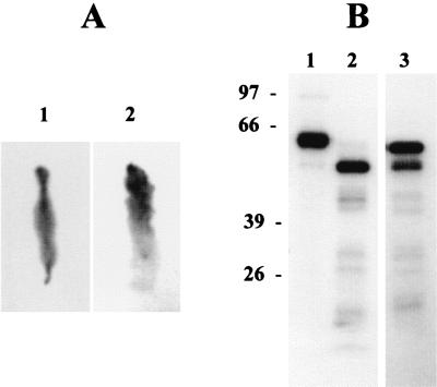
FIG. 2.
Autoradiography of L. dispar larval midgut and digested Cry2Aa1 after 1 h of feeding on diet contaminated with 125I-labeled Cry2Aa1. (A) The peritrophic membrane with its contents (image 1) and midgut tissue after removal of peritrophic membrane (image 2). Note that the anterior regions of both the peritrophic sac and the midgut tissue were arranged upward. (B) Lanes: 1, 125I-labeled Cry2Aa1 before it was applied to the insect; 2, 125I-labeled Cry2Aa1 extracted from the midgut fluid; 3, 125I-labeled Cry2Aa1 extracted from the midgut membrane of the same insect as in lane 2.

