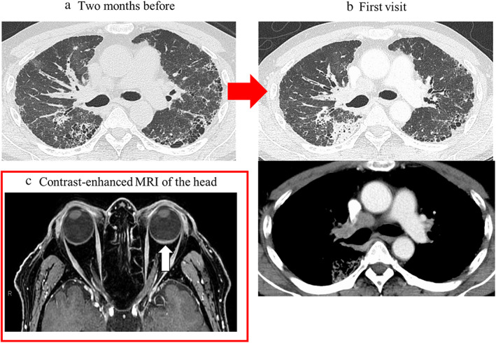
FIGURE 1.

(a) Computed tomography (CT) scan image acquired 2 months prior to the first visit to our department and (b) at the time of the first visit to our department (b). (a) Reticular shadows and cystic clusters with a predominance of subpleural areas in bilateral lungs as ADM‐associated interstitial lung disease are seen. (b) New mass shadow in the right upper lobe approximately 3.0 cm in length and diameter and mediastinal lymphadenopathies are newly observed. (c) Contrast‐enhanced magnetic resonance imaging of the head, showing left retinal thickening (white arrow). Abbreviations: ADM: amyopathic dermatomyositis; CT: computed tomography; MR: magnetic resonance imaging
