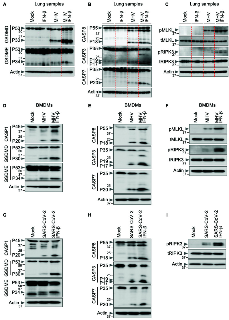
Fig. 3. IFN-β promotes inflammatory cell death, PANoptosis, during β-coronavirus infection.

(A–C) Immunoblot analysis of (A) pro- (P53) and activated (P30) gasdermin D (GSDMD), pro- (P53) and activated (P34) gasdermin E (GSDME); (B) pro- (P55) and cleaved caspase-8 (CASP8; P18), pro- (P35) and cleaved caspase-3 (CASP3; P19 and P17) and pro- (P35) and cleaved caspase-7 (CASP7; P20); and (C) phosphorylated MLKL (pMLKL), total MLKL (tMLKL), phosphorylated RIPK3 (pRIPK3) and total RIPK3 (tRIPK3) in the lung samples from mock- or IFN-β–treated wild type (WT) mice with or without mouse hepatitis virus (MHV) infection 3 days post-infection. (D–I) Immunoblot analysis of (D, G) pro- (P45) and activated (P20) caspase-1 (CASP1), pro- (P53) and activated (P30) GSDMD, pro- (P53) and activated (P34) GSDME; (E, H) pro- (P55) and cleaved CASP8 (P18), pro- (P35) and cleaved CASP3 (P19 and P17) and pro- (P35) and cleaved CASP7 (P20); (F) pMLKL, tMLKL, pRIPK3 and tRIPK3; and (I) pRIPK3 and tRIPK3 in mock- or IFN-β–treated bone marrow-derived macrophages (BMDMs) or THP-1 cells during MHV or SARS-CoV-2 infection, respectively. Actin was used as the internal control. Molecular weight marker sizes in kDa are indicated in small font on the left of each blot. Asterisk denotes non-specific bands (A, G). Data are representative of at least three independent experiments.
