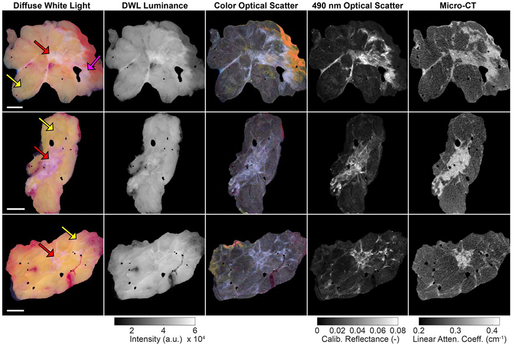
Figure 1.

Wide field-of-view images of representative invasive ductal carcinoma specimens (yellow arrows = adipose tissue; pink arrow = connective tissue; red arrows = malignant tissue). 1 cm scale bars are shown in the first column. In the optical images, surgical ink (yellow, orange, red) is visible along some specimen margins. Linear attenuation coefficient values correspond to 50 kVp. DWL = diffuse white light.
