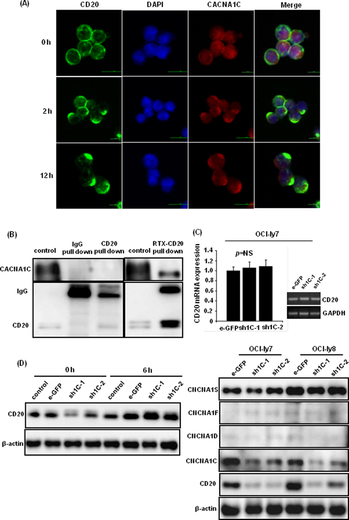
Figure 3: The interaction between CACNA1C and CD20 molecules during rituximab action.

(A) CD20 (green light) and CACNA1C (red light) molecules exhibited a similar polarized distribution after rituximab treatment of 0, 2, and 12 h. Their overlapping distribution (yellow light) was captured in the plasma membrane in the merged phase. (B) CACNA1C protein was detected in anti-CD20 pull-down protein using western blot after rituximab treatment of 2 h in OCI-ly7 cells. (C) Knockdown of CACNA1C expression with transduced shRNAs did not affect CD20 mRNA levels, but downregulated CD20 protein expression. Other isoforms of α1 subunits, such as CACNA1D, CACNA1F, and CACNA1S, showed no obvious alteration. (D) Proteasome inhibitor bortezomib (20nM) restored CD20 expression after administration of 6 h in OCI-ly7 cells.
