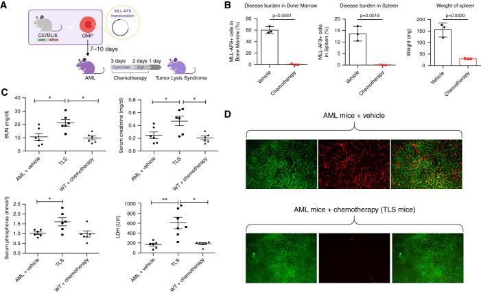
Figure 1.
Biochemical and microscopic analysis of a mouse model of AML mice show significant TLS after administration of chemotherapy. (A) Model of TLS in AML mice. (B) Disease (percent MLL-AF9+ blasts) in the bone marrow and spleen of MLL-AF9 mice after treatment with vehicle or with chemotherapy (24 hours after treatment). n=3 per group. (C) BUN, LDH, serum creatinine, and phosphate were measured in AML mice treated with vehicle (n=6) or chemotherapy (cytarabine + doxorubicin; n=6) and WT mice treated with chemotherapy (n=6) 24 hours after treatment. (D) In vivo renal intravital microscopy in MLL-AF9 mice that received vehicle or chemotherapy. MLL-AF9 leukemic blasts are DsRED positive and visible in renal peritubular capillaries. After chemotherapy, blast cell infiltration is no longer visible. All data are presented as mean±SEM *P<0.05, **P<0.01 versus vehicle, Mann–Whitney test.

