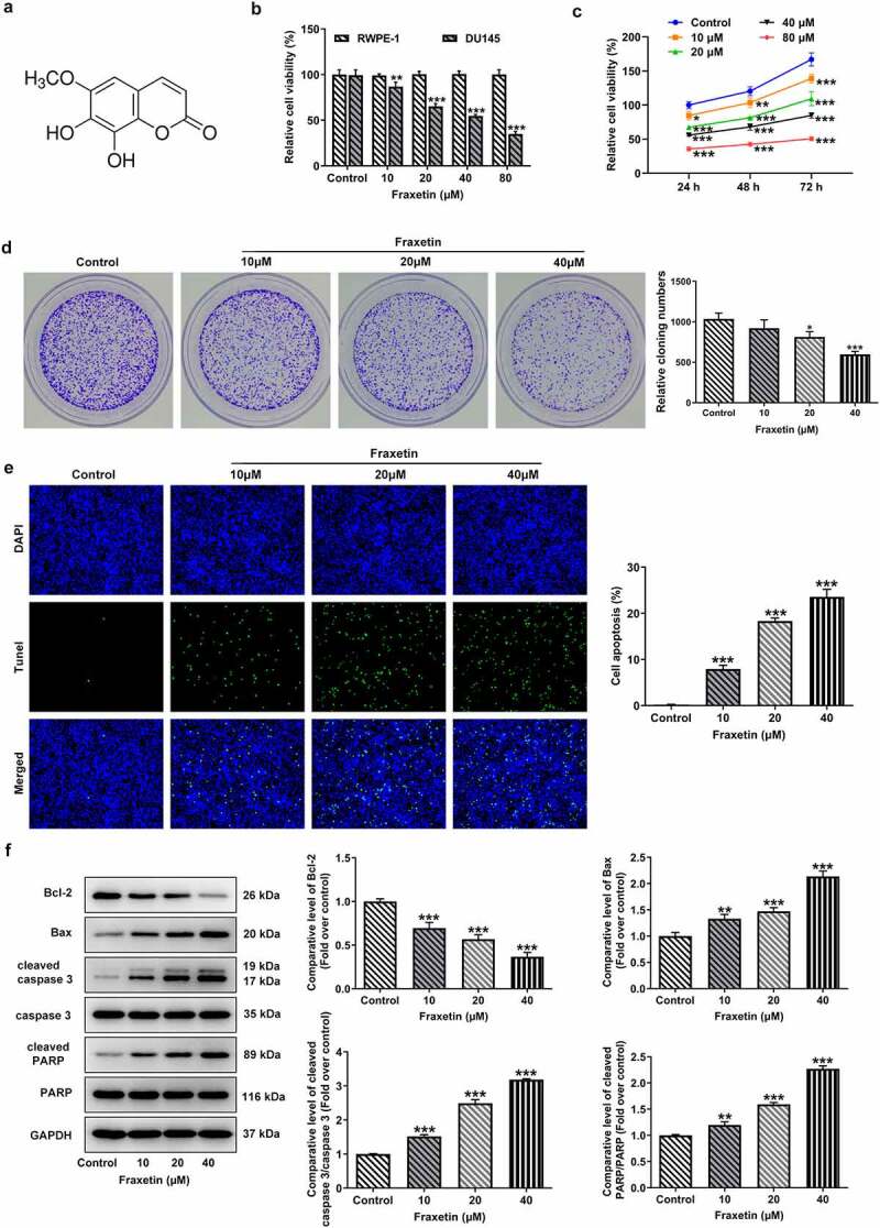
Figure 1.

Fraxetin inhibits proliferation and induces apoptosis of DU145 prostate cancer cells. (a) the chemical structure of Fraxetin. (b) RWPE-1 and DU145 cells were treated with 0, 10, 20, 40 and 80 μM Fraxetin for 48 h, then cell viability was assessed using CCK-8 assay. (c) DU145 cell was treated with to 0, 10, 20, 40 and 80 μM Fraxetin for 24, 48 and 72 h, then cell viability was detected using CCK-8 assay. (d) colony formation assay was used to detect proliferation of DU145 cells that in the presence of 0, 10, 20 and 40 μM Fraxetin. E, the apoptosis of DU145 cell was observed by Tunel staining (magnification, x200). F, the protein expression of Bcl-2, Bax, cleaved-caspase 3/caspase 3 and cleaved PARP/PARP in DU145 was detected by western blot assay. *P < 0.05, **P < 0.01 and ***P < 0.001 vs Control.
