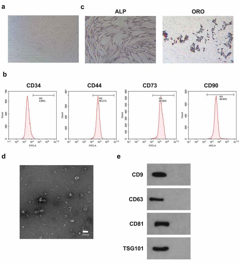
Figure 5.

Identification of BMSCs and Exo.
A. Observation of the morphology of BMSCs with an inverted microscope; B. Test of BMSCs surface markers CD44, CD73, CD34 and CD90 was via flow cytometry; C. ALP staining and Oil-Red-O staining to Examination of ALP activity and lipid after BMSCs osteogenic or adipogenic induction was via ALP staining and Oil-Red-O staining; D. Observation of Exo morphology was via TEM; E. Test of Exo-labeled proteins CD9, CD63, CD81 and TSG101 was via Western blot.
