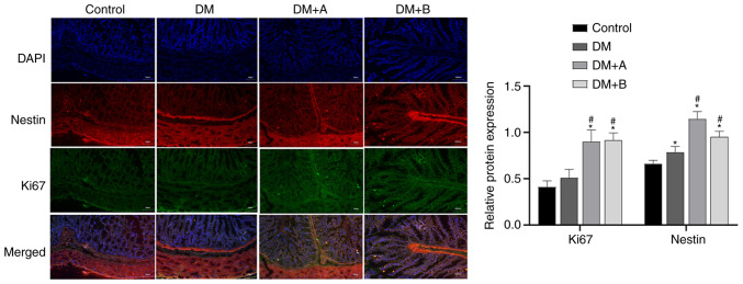
Figure 6.
Immunofluorescence double staining images of Nestin and Ki67 in the Control, DM, DM + A and DM + B groups. DAPI-stained nuclei appeared blue. Nestin and Ki67 are indicated by red fluorescence and green fluorescence, respectively. A rat model of DM was established using an intraperitoneal injection of streptozotocin. The rats in the Control group were given an equal volume of citric acid solvent. Magnification, ×100. *P<0.05 vs. Control; #P<0.05 vs. DM. DM, diabetes mellitus model; DM + A, diabetic rats treated with 5 µg/kg prucalopride; DM + B, diabetic rats treated with 10 µg/kg prucalopride.

