
Fig. 1.
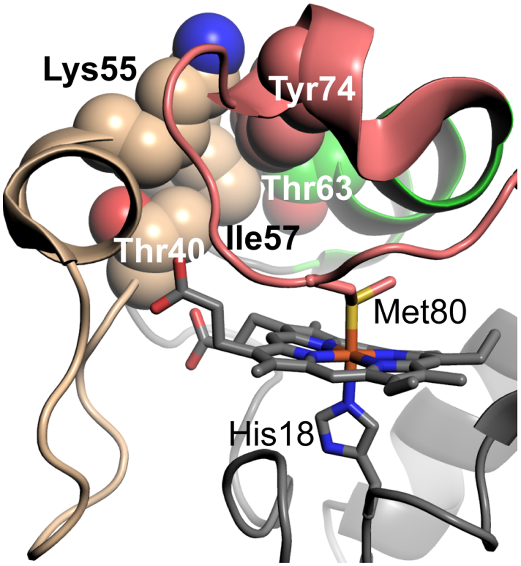
Visualization of human Cytc (PDB ID: 3ZCF) [26] residues (space-filling models) in or near Ω-loop C (shown in wheat) that have coevolved. Ω-loop D is shown in salmon and the 60s helix in green. Shown as stick models are the heme and its axial ligands Met 80 and His 18.