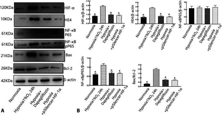
FIGURE 5.

Dapagliflozin protected tubular epithelial cells through the HIF-1α/HE4/NF-κB signaling pathway. A, Western blot analysis of the expression of HIF-1α, HE4, P65, and pP65 in HK-2 cells treated with dapagliflozin and incubated hypoxia for 24 hours. Hypoxia increased the expression of Bax, whereas dapagliflozin decreased those of Bax and increased those of Bcl-2, respectively, in HK-2 cells. B, Representative quantitative analysis. *P< 0.01 versus the normoxia group; & P < 0.05 versus the dapagliflozin + hypoxia group. #P < 0.05 versus the hypoxia + pSilencer-HIF-1α group. There are 3 replicates in each group for cellular experiments.
