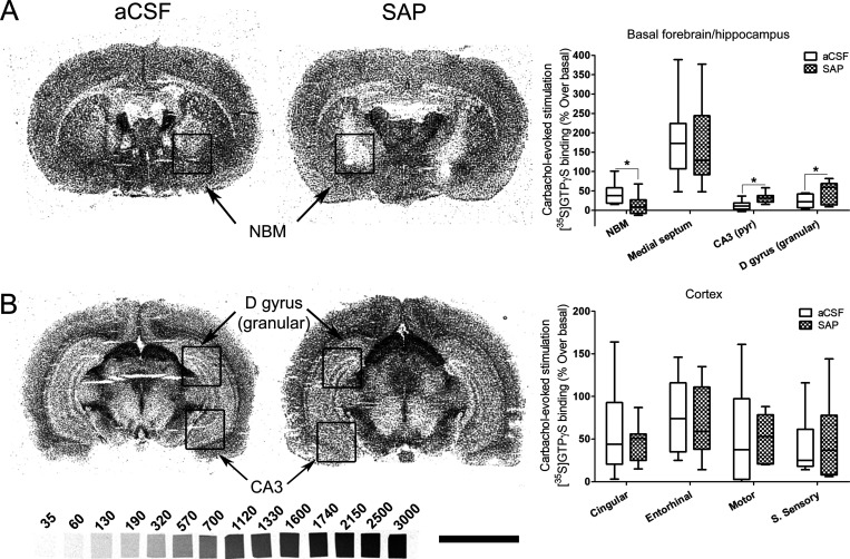
Figure 5.
[35S]GTPγS autoradiography in rat brain coronal sections at two different levels from Bregma, (A) including NBM and (B) including dorsal hippocampus, obtained from aCSF, left (n = 9), and SAP-treated rats, right (n = 11), that show representative autoradiograms of [35S]GTPγS binding evoked by carbachol (100 μM). This assay is specific to Gi/o coupled receptors; therefore we are measuring the activity mediated by M2/M4 mAChR. The graphs show the mean ± SEM of each group in the different analyzed areas. NBM: nucleus basalis magnocellularis. D gyrus (granular): granular dentate gyrus. CA3: CA3 region of hippocampus. S sensory: somatosensory cortex. [14C]-Microscales were used as standards in nCi/g t.e. Scale bar: 5 mm.

