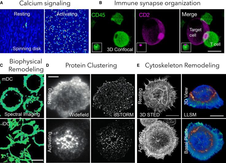
Figure 2. Potential applications of different imaging modalities in immunity.
(A) Calcium signaling with spinning disk microscopy [116] where bright spots show calcium flux (scale bar 20 µm); (B) 3D immune synapse with confocal microscopy [146] where magenta is CD2 on target synthetic cells and green is CD45 in T cells (scale bar 10 µm); (C) biophysical imaging with spectral imaging combined with smart probes [103] where immature dendritic cells (iDCs) show lower membrane fluidity than mature dendritic cells (mDCs) (scale bar 10 µm); (D) protein clustering with SMLM [48] (scale bar 3 µm); (E) cytoskeleton imaging with STED and LLSM [33] (scale bar 10 µm).

