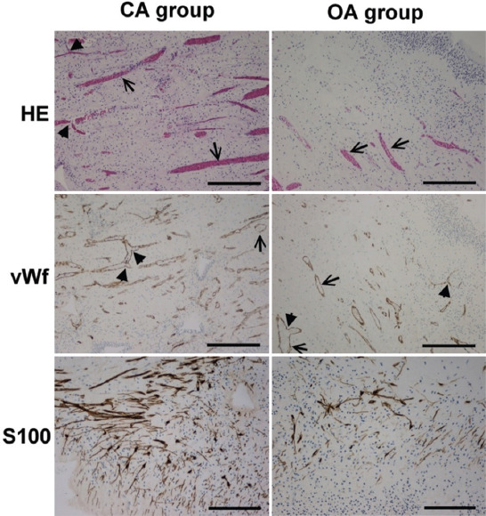
FIGURE 3.

Crown part of dental pulp with closed (CA group) and open apex (OA group) stained with HE (objective magnification ×4, bar=300μm), anti-vWf (objective magnification ×4, bar=300μm), and anti-S100 (sub-odontoblast zone, objective magnification ×20, bar=150μm). Note that the pulp tissue is insignificantly more vascularized (HE, vWf) and significantly more innervated (S100) in the CA group than in the OA group. ↑: Vessels with the thin wall; ▲: Branching of the vessels in the right angels.
