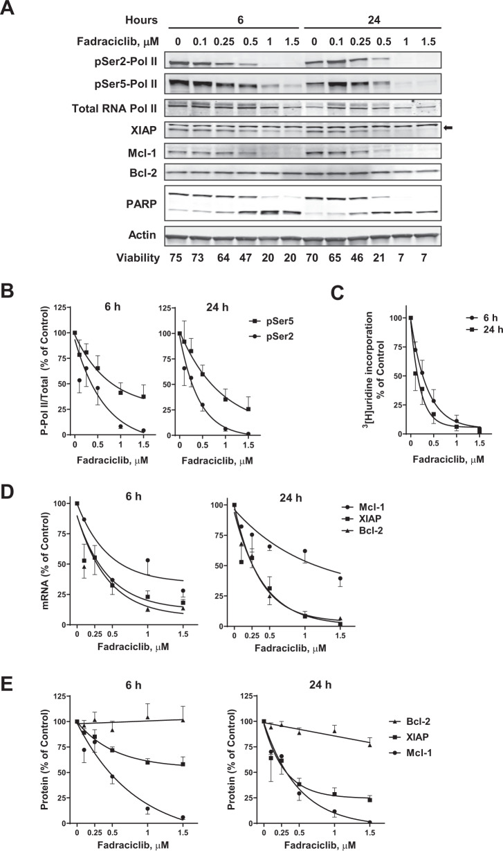
Fig. 1. Mechanism of action of fadraciclib in primary CLL cells.
A Fadraciclib reduced the phosphorylation of RNA Pol II and reduced the anti-apoptotic proteins Mcl-1 and XIAP. CLL cells were incubated with increasing concentrations of fadraciclib for 6 and 24 h, and the phosphorylation of RNA Pol II was analyzed by immunoblotting, using antibodies against the phosphorylated Ser2 or Ser5 sites of the CTD, as well as total Pol II. The major anti-apoptotic proteins Mcl-1, Bcl-2 and XIAP were analyzed with their specific antibodies. PARP was used as an indicator of apoptosis, and actin was used as a loading control. A representative immunoblot of 8 patient samples is shown. Cell viability, measured by annexin V/propidium iodide (PI) and flow cytometry, is shown below the bands. Arrow at right indicates the band for XIAP. B Inhibition of phosphorylation of Pol II at Ser2 (●) and Ser5 (■) sites after 6 h (left) and 24 h (right) incubation with fadraciclib. Levels of phosphorylation were quantified from the blots in (A), normalized to total Pol II, and then expressed as a percentage of time-matched controls (mean ± standard error of the mean [SE] of 8 individual CLL samples). C Inhibition of RNA synthesis by fadraciclib in CLL cells. RNA synthesis was measured by [3H]uridine incorporation in 5 CLL samples after 6 (●) and 24 h (■) incubation with increasing concentrations of fadraciclib. Each measurement was performed in triplicate. Data are presented as percentage of time-matched controls (mean ± SE of 5 CLL samples). D Fadraciclib reduced mRNA levels of Mcl-1, XIAP, and Bcl-2. mRNA levels of Mcl-1(●), XIAP (■), and Bcl-2 (▲) were measured by real-time RT-PCR, each performed in duplicate, and compared with time-matched controls (mean ± SE of 8 CLL samples). E Quantitation of immunoblots of Mcl-1, XIAP, and Bcl-2 from the same samples as described in (A). Levels of Mcl-1, XIAP, and Bcl-2 were normalized to actin and expressed as percentage of time-matched controls (mean ± SE of 8 CLL samples).

