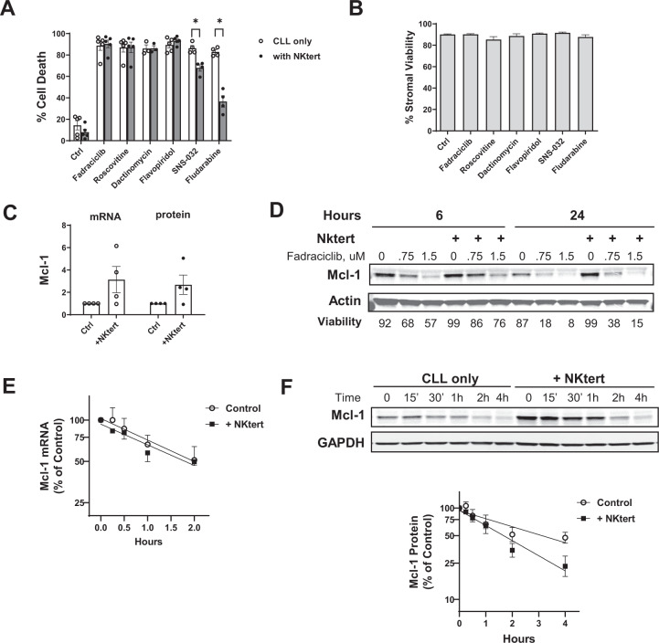
Fig. 3. Fadraciclib overcame protection conferred by stroma cells.
A CLL cells were incubated with approximately 2× IC50 concentrations of each compound without (CLL only) or with a layer of StromaNKtert cells (+NKtert) for 24 h, except for fludarabine (48 h). Cell death were measured in the CLL cells by annexin/PI staining and presented as mean ± SE of 5 CLL samples. A paired t-test was performed comparing cell death induced by each compound in the “CLL only” group to the “NKtert” group. The differences were not significant except for SNS-032 and fludarabine (noted by the stars above the bars). B The compounds used in (A) were not toxic to the StromaNKtert cells. The StromaNKtert cells were exposed to the same concentrations of the compounds at the same conditions as in (A), and the viability of the stroma cells was measured by annexin/PI staining. Data represent viability (mean ± SD) of measurements performed in triplicates. C Co-incubation of the CLL cells with the stroma cells induced Mcl-1 mRNA and protein expression. The CLL cells were incubated with or without the stroma layer for 24 h. The mRNA level of Mcl-1 was measured by real-time RT-PCR, and the protein level was measured by immunoblotting; each was expressed as the ratio to the levels of the controls in CLL cells cultured alone (mean ± SE of 4 CLL samples). D CLL cells reduced Mcl-1 level in the presence of StromaNKtert cells. CLL cells were incubated in the absence or presence of stroma cells at increasing concentrations of fadraciclib for 6 and 24 h. Mcl-1 levels were determined by immunoblotting. One immunoblot that is representative of those obtained from 3 patient samples is shown, and cell viabilities are displayed below the image. E Mcl-1 mRNA half-life in the presence or absence of stroma cells. CLL cells were incubated with 5 μg/ml dactinomycin in the presence or absence of stroma cells. The cells were collected at 0, 0.25, 0.5, 1, 2, and 4 h, and the Mcl-1 mRNA level was measured by real-time RT-PCR and plotted as percentage of Mcl-1 level over the controls (mean ± SE of 4 samples). F The half-life of Mcl-1 protein in the absence and presence of the stroma cells. CLL cells were incubated with 50 μg/ml cycloheximide in the presence and absence of stroma cells. Cell pellets were collected at indicated times, and Mcl-1 levels were measured by immunoblotting. In all samples, Z-VAD-FMK (50 μM) was added to prevent loss of Mcl-1 due to caspase cleavage. A representative immunoblot from 5 experiments is shown, and the protein levels were quantified and plotted as percentage of Mcl-1 level/controls (mean ± SE of 5 CLL samples).

