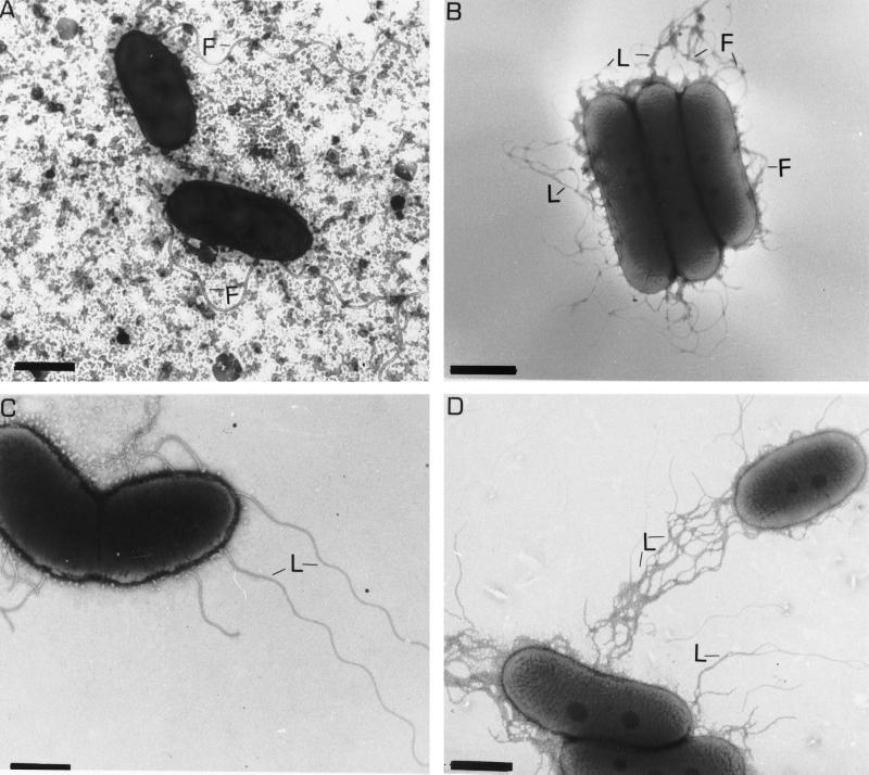
FIG. 1.
Transmission electron micrographs of A. lipoferum 4B (A and B) and 4VI (C and D) cells. Cells were grown in liquid (A and C) and on solid (B and D) media. The lateral flagella are thinner in diameter and have a shorter wavelength. Abbreviations: F, polar flagellum; L, lateral flagella. Bars, 1 μm (A, B, and D) and 0.7 μm (C).

