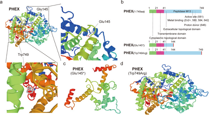
Fig. 6. The structure prediction map of PHEX protein mutant.
a three-dimensional crystal structure map of PHEX protein and the location of mutation sites (dmam SwissMe model database, URL: https://swissmodel.expasy.org/repository/uniprot/P78562). b the functional domain map of PHEX protein and its variants. c, d prediction and comparison of the three-dimensional structure of normal PHEX and Glu145* mutation and Trp749Arg mutation using Swiss-model. (URL: https://swissmodel.expasy.org/repository/uniprot/P78562).

