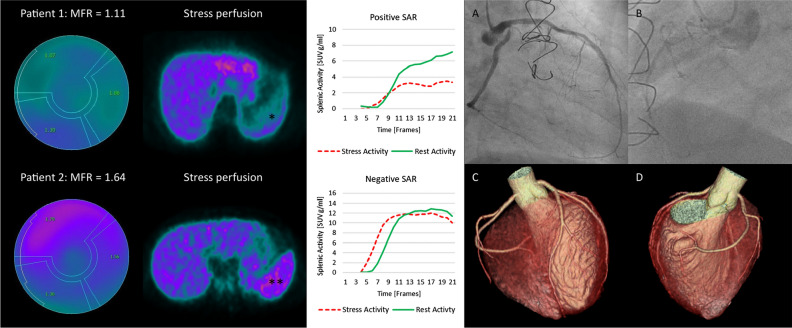
Fig. 7.
An example of two patients with impaired MFR. Invasive coronary angiography of patient #1 (top) with impaired MFR of 1.11 and a SAR of 0.41 (positive SSO: *) revealed severe CAD with a subtotal stenosis of the ostial left circumflex artery (A), occluded RCA (B) and subtotal stenosis of LIMA-LAD bypass graft, indicating truly reduced MFR. Coronary computed tomography (C,D) in patient #2 (bottom) with impaired MFR of 1.64 and a SAR of 0.93 (negative SSO: **) excluded obstructive CAD or calcifications, indicating failed adenosine response as the cause for the impaired MFR

