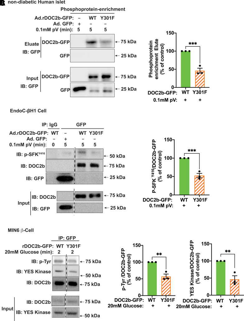
Figure 6.
Y301F mutation reduces the tyrosine phosphorylation of DOC2b and blocks its interaction with activated SFK and YES in the β-cell. A, left: Representative Western blot images of nondiabetic human islets transduced with Ad.GFP, Ad.rDOC2b-GFPWT, or Ad.rDOC2b-GFPY301F, incubated for 48 h, and treated with pV (0.1 mmol/L, 5 min) followed by phosphoprotein enrichment and immunoblot (IB) analysis. A, right: Quantification of the IBs from three independent experiments. B, left: Representative IB images of human EndoC-βH1 cells transduced with Ad.GFP, Ad.rDOC2b-GFPWT, or Ad.rDOC2b-GFPY301F and treated with or without 0.1 mmol/L pV for 5 min followed by IP and IB analysis. B, right: Quantification of the IBs from three independent experiments. C, left: Representative Western blot images of MIN6 β-cells transfected with either rDOC2b-GFPWT or rDOC2b-GFPY301F and stimulated with or without glucose for 2 min followed by IP and IB analysis. Vertical dashed lines denote splicing of lanes from within the same gel exposure. C, right: Quantification of the IBs from three independent experiments. Data are shown as mean ± SEM. **P < 0.002; ***P < 0.0002.

