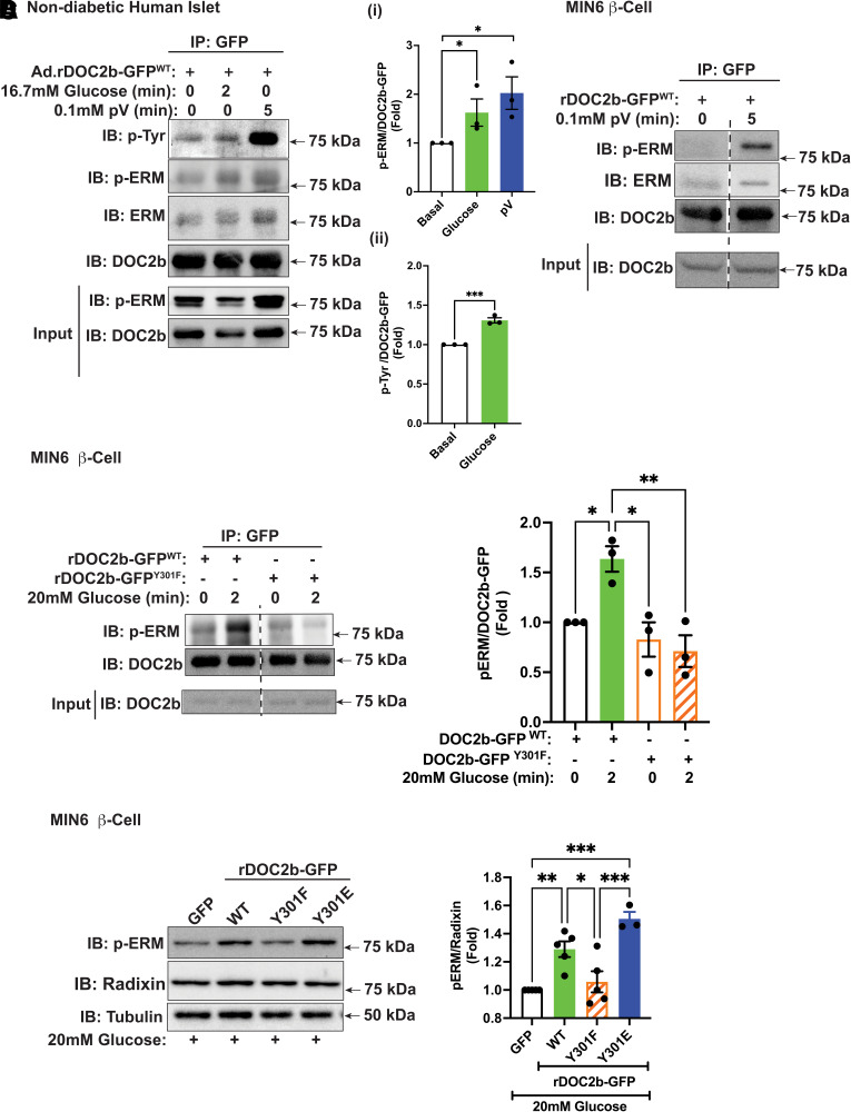
Figure 8.
Glucose stimulation and pV treatment augment the binding of DOC2b with p-ERM in β-cells. A: Representative Western blot images of nondiabetic human islets transduced with Ad.rDOC2b-GFPWT for 48 h and treated with or without 20 mmol/L glucose (2 min) or 0.1 mmol/L pV (5 min) followed by IP and immunoblot (IB) analysis. Ai: p-ERM/DOC2b ratio, quantified from three independent sets of experiments. Aii: pTyr/DOC2b ratio, quantified from three independent experiments. B: Representative Western blot images of MIN6 β-cells transfected with rDOC2b-GFPWT for 48 h and treated with or without 0.1 mmol/L pV (5 min), followed by IP and IB analysis. Images are representative of three independent experiments using independent passages of cells. C, left: Representative IB images of MIN6 β-cells transfected with rDOC2b-GFPWT or rDOC2b-GFPY301F for 48 h and treated with or without 20 mmol/L glucose (2 min) followed by IP and IB analysis. Bar graph quantification of three independent experiments shown at right. D, left: Representative images of MIN6 β-cells transfected with GFP or rDOC2b-GFPWT or rDOC2b-GFPY301F or rDOC2b-GFPY301E for 48 h and treated with 20 mmol/L glucose for 30 min followed by IB analysis. Bar graph quantification of the IBs from three independent experiments shown at right. Anti-tubulin antibody was used as a loading control. Data are shown as mean ± SEM. *P < 0.05; **P < 0.002; ***P < 0.0002.

